2020/04 The World-first Research Finding Shows Evidence for Fossil Fuel PM1 Accumulation in Marine Biota
The World-first Research Finding Shows Evidence for Fossil Fuel PM1 Accumulation in Marine Biota
Professor Li-Lian Liu led a research team from the Department of Oceanography published a world-first research finding on the world-leading journal, Environmental Science & Technology 2020 54 (7), 4068-4078 recently. Their finding shows evidence for PM1 that released from fossil fuel would accumulate in marine biota when they enter the ocean. The research result resolved the inherent technical difficulties in obtaining particulates from biological samples for qualitative and quantitative characterization.
When fine particulates such as those with a diameter of approximately 1 μm (particulate matter, PM1) are released from fossil fuel combustion into the air, they warm the atmosphere and contribute to millions of premature deaths in humans each year. Considerable quantities of PM1 eventually enter the oceans as suspended particulates, yet subsequent removal mechanisms are poorly understood. In fact, the presence of PM1 in marine biota has never been reported. Since sea anemones are opportunistic suspension feeders, they are anticipated to incorporate and accumulate PM1 in their bodies. By histological examination, PM1 was detected in 21 of the 22 sea anemones collected from Taiwan and Southeast China, with a depth of intertidal zone to 1000 m. PM1, if present, was always detected in endodermal layers and had the same dominant color (i.e., black, brown, or green) in different species from the same site. The bioaccumulation factor of PM1 in sea anemones was approximately 5−7 orders of magnitude. Based on radioisotope 14C results, the contribution of fossil fuel source PM1 was 8−24%. Regardless of PM1’s color, S and Fe were commonly detected by scanning electron microscopy and energy-dispersive spectrometry (SEM-EDS), suggesting anthropogenic sources. Furthermore, a maternal transfer of materials was suggested based on the existence of PM1 in sea anemone eggs and in brooding and released juveniles. The significance of PM1 accumulation by biota in aquatic ecosystems and the potential risk to living organisms via food webs warrant further investigation.
Professor Liu’s team included Chair Professor, Chen-Tung Arthur Chen, Assistant Professor, Hon-Kit Lui, and two students Chen-Yun Hsieh, Meng-Ying Kuo of the Department of Oceanography. Besides, Associate Professor, Yen-Hong Shau of the Department of Marine Biotechnology and Resources, Professor Chung-Shin Yuan from the Institute of Environmental Engineering, and Dr. Chienhsun Chen, an associate researcher at Taiwan Ocean Research Institute are joint members. The team has recently focused on the effects of PM on biological systems and the contribution of biota-PM to the black cycle in oceans.
Paper Link to Environmental Science & Technology
https://dx.doi.org/10.1021/acs.est.9b06976
Contact:
Professor Li-Lian Liu,
Department of Oceanography, National Sun Yat-sen University
TEL: 886-7-5252000*5108
Email: lilian@mail.nsysu.edu.tw

Figure 1. The particulate matter (PM) in sea anemone bodies. (A) Anthopleura sp. from Kuishantao Islet collected in June 2018. (B) PM in a tentacle; (C) enlarge image of (B); (D) Diadumene lineata from Taichung Harbour collected in July 2018; (E) PM in a tentacle of D. lineata from Hanbao collected in July 2018; (F) enlarge image of (E). Red arrow: PM; ec: ectoderm; en: endoderm; m: mesoglea.
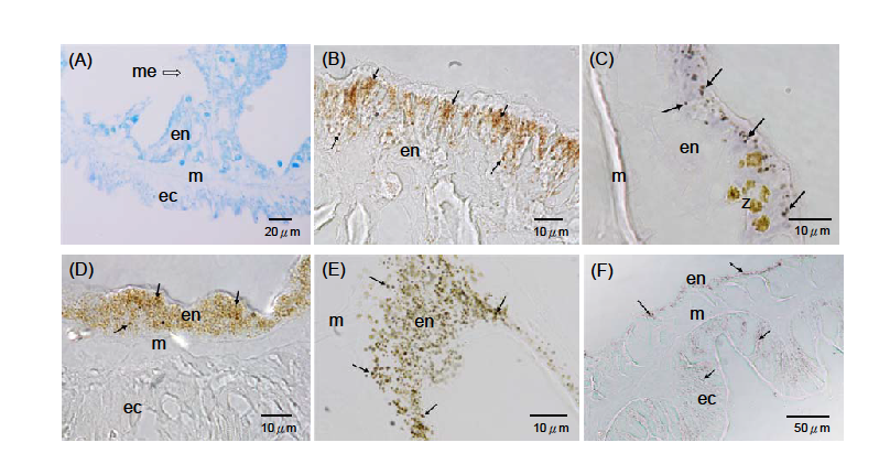
Figure 2. Photomicrographs showing particulate matter (PM) with different dominant colors and sizes detected in various sea anemones near Taiwan. (A) No PM in Boloceroides mcmurrichi sampled at the Dongsha Island intertidal zone in Jan. 2016; (B) brown PM in Stylobates aeneus endoderm sampled off the Xiaoliuqui Island at a depth of 1000 m in July 2008; (C) black PM in Anthopleura asiatica endoderm sampled at the Magang intertidal zone in March 2017; (D) brown PM in Diadumene lineata endoderm sampled at the Hanbao intertidal zone in Feb. 2000; (E) green PM in D. lineata endoderm sampled at the Mailiao intertidal zone in March 2018; (F) black PM in Anthopleura sp.2 endoderm and ectoderm sampled at the Shimen intertidal zone in Sept. 2016. →: PM; ec: ectoderm; en: endoderm; m: mesoglea; me: mesentery; z: zooxanthellae.
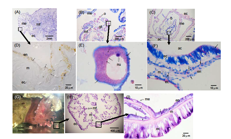
Figure 3. Particulate matter (PM) observed in reproductive sea anemones and offspring. (A) Longitudinal fission progeny of Anthopleura asiatica sampled at Magang in March 2017; (B) mature oocytes of Diadumene lineata sampled at Xiangshan in April 1999; (C) brooding juvenile of Anthopleura sp.1 sampled at Huxia in Sept. 2017; (D) enlarged image of (A); (E) enlarged image of (B); (F) enlarged image of (C); (G) parent with hatched juveniles of Actinia equina sampled at Magang in March 2016; (H) cross section of an A. equina juvenile; (I) enlarged image of (H). →: PM; ac: actinopharynx; b: brooded juvenile; ec: ectoderm; en: endoderm; gt: gametogenic tissue; m: mesoglea; me: mesentery; mf: mesenterial filament; nu: nucleolus; o: oocyte; z: zooxanthellae.